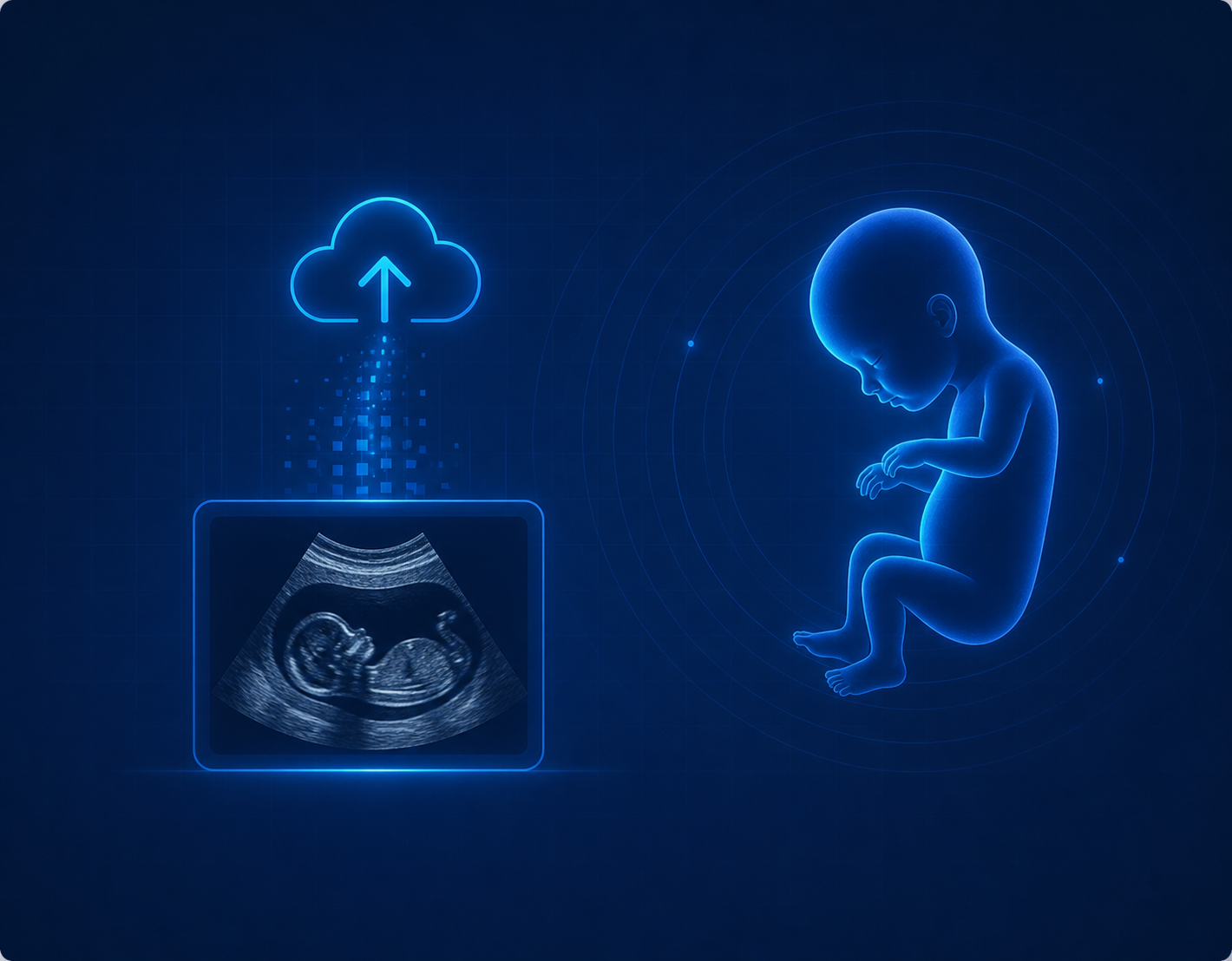
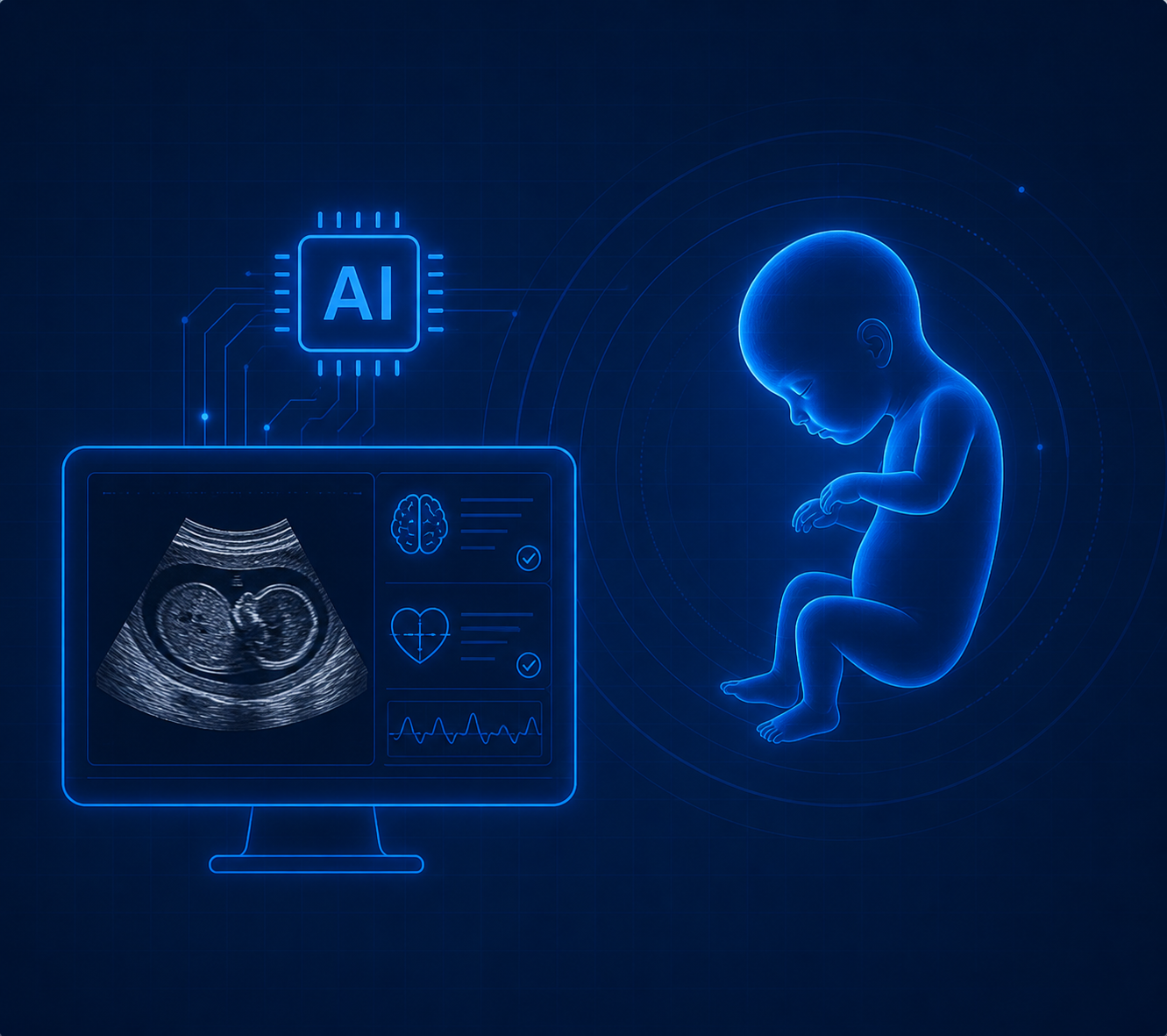
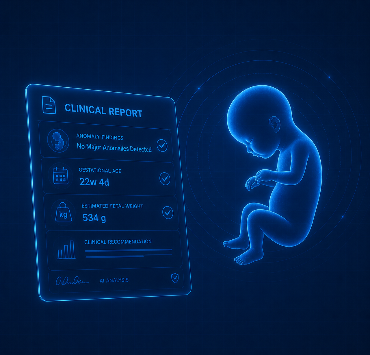
Using AI trained on 3,000+ annotated fetal brain ultrasound images to detect abnormalities in the CSP and Lateral Ventricles — enabling early, accurate prenatal diagnosis.
The FADA: Fetal Abnormality Detection Algorithm project is led by the research team at Hamad Bin Khalifa University (HBKU) in Qatar, supported by the Global Health Institute (GHI) at the American University of Beirut (AUB).
Funded by the Canadian International Development Research Centre (IDRC) and the UK International Development (FCDO), FADA has annotated over 3,000 fetal brain ultrasound images — building an expert-labelled dataset focused on the Fetal Brain, CSP (Cavum Septum Pellucidum — a small fluid-filled space in the brain; its absence signals serious development problems), and LV (Lateral Ventricles — fluid chambers deep in the brain; enlargement beyond 10 mm can indicate ventriculomegaly, a sign of abnormal brain growth).
Our intelligent platform transforms the prenatal diagnostic process into a simple, fast, three-step workflow.
Securely upload a fetal brain ultrasound scan. FADA accepts standard scan images taken during routine prenatal check-ups.
Our model — trained on 3,000+ expert-annotated images — detects the CSP (a fluid space whose absence flags brain defects) and measures LV width (enlargement above 10 mm indicates ventriculomegaly).
Receive a detailed report with anomaly findings, gestational age estimate, fetal weight, and evidence-based clinical recommendations for the doctor.
FADA is trained on 3,000+ expert-annotated fetal brain ultrasound images to identify three key structures. Here is what each one means.
The Developing Brain
The brain of a baby still in the womb. During pregnancy, certain structures inside the fetal brain can be seen clearly on an ultrasound scan. FADA checks these structures to spot signs of abnormal development as early as possible — giving doctors time to plan the safest care.
Normal range: 3 – 10 mm
A small, fluid-filled space located in the centre of the brain, between two thin sheets of tissue. Think of it as a tiny gap that should always be visible on a mid-pregnancy scan. If it is absent, it can indicate serious brain development problems such as holoprosencephaly (failure of the brain to divide into two halves) or septo-optic dysplasia (underdevelopment of key brain structures).
Normal: ≤ 10 mm width
Two fluid-filled chambers deep inside the brain that cushion and nourish it. They are like the brain's internal water balloons. When they grow wider than 10 mm, the condition is called ventriculomegaly — meaning the ventricles are abnormally enlarged. Mild cases often resolve on their own; severe enlargement can signal conditions such as hydrocephalus (excess fluid build-up) or chromosomal abnormalities.
FADA is driven by three core objectives that shape our research and clinical deployment strategy.
Empower healthcare professionals with AI-assisted decision-making tools that augment clinical expertise and improve diagnostic accuracy.
Support more accurate and timely detection of potential fetal anomalies, significantly reducing the risk of missed or delayed diagnoses.
Improve health outcomes for both mother and baby by enabling earlier, more informed, and evidence-based clinical interventions.
A multidisciplinary team of researchers, professors, AI experts, and developers from Hamad Bin Khalifa University, Qatar.
Post Doc Fellow (Lead PI)
HBKU, Qatar
Professor
HBKU, Qatar
Professor
HBKU, Qatar
AI Expert
HBKU, Qatar
Software Developer
HBKU, QatarJoin our platform and leverage AI-assisted fetal anomaly detection to improve outcomes for mothers and babies.